In-Depth Exploration of Head and Neck Anatomy


Intro
Understanding the anatomy of the head and neck is crucial for a wide range of fields—from medical education to artistic endeavors. This intricate region houses vital structures that play a significant role in the functioning of the body. From the delicate arrangement of facial muscles to the complex system of nerves and blood vessels, every detail matters.
With a robust foundation in the anatomical features, students, educators, and professionals are better equipped to grasp not just anatomy but also the interplay between form and function. These insights can contribute to advancements in healthcare, improve artistic representations in media, and enhance educational approaches.
"To appreciate the beauty of the head and neck’s anatomy is to understand its function; both aspects are inseparable."
This article endeavors to present a well-rounded view of the head and neck, focusing not on just key components, but also on how they interact. The aim is simple yet profound—bridging the conceptual divide between complex anatomical frameworks and everyday applications.
Preface to Head and Neck Anatomy
Understanding the anatomy of the head and neck is no small feat; it’s like piecing together a complex puzzle where each piece plays an instrumental role in both structure and function. The head and neck region houses an array of vital systems, including the respiratory, digestive, and sensory systems, intertwining to support basic functions like breathing, swallowing, and expressing emotions.
This article intends to provide a thorough exploration of this intricate area. By laying bare the underlying anatomical framework, it aims to elevate the reader's comprehension. This understanding is particularly crucial for students, researchers, and healthcare professionals who rely on precise knowledge of anatomy in their respective fields. Grasping these intricate relationships can lead to better diagnostic and therapeutic strategies.
Overview of Anatomical Significance
The significance of head and neck anatomy extends beyond the confines of academia; it resonates within everyday life. For instance, consider the delicate interplay between the facial muscles and our ability to communicate non-verbally. Each expression carries meaning, shaped by anatomical intricacies like the temporalis and masseter muscles working in tandem to facilitate chewing and facial movement.
Moreover, structures such as the larynx play a pivotal role not just in phonation, but also in protecting the airway. When understanding the layout of these structures, professionals can diagnose and treat disorders more effectively. Without a minute knowledge of this region, one might overlook conditions such as laryngeal cancer, which demands early detection.
Purpose and Scope of the Article
The purpose of this article goes beyond providing visuals and descriptions; it seeks to forge a connection between theoretical knowledge and clinical applications. As we navigate through different sections, readers will encounter comprehensive discussions on skull structure, muscular anatomy, vascular systems, and more. Each segment is meticulously crafted to ensure clarity and depth.
The scope of this article encompasses not only the anatomical figures and isolated structures but also their interdependence.
- We will delve into how cranial nerves influence functions ranging from sensation to motor control.
- The implications of the vascular network on the overall health of the head and neck will also be explored.
By the end, readers will have a well-rounded understanding of how these components foster harmony and function in a dynamic environment. This will serve not just as a reference but as a roadmap to deeper anatomical exploration.
Skull Structure
The skull is a remarkable and complex structure that serves various roles in both anatomical and functional contexts. The arrangement of the skull not only provides a protective casing for the brain but also establishes the framework for multiple sensory organs and facilitates crucial connections for various physiological functions. Understanding the skull structure is vital in fields ranging from medicine to anthropology, as it sheds light on the evolutionary adaptations of humans and other species.
In discussing the skull, we can break it down into three primary components: the cranial bones, the facial bones, and the sutures and joints that bind them together. Each element reveals profound details about human biology and its interconnections with external factors like environment and growth patterns.
Cranial Bones
The cranial bones consist of eight individual bones that come together to form the cranial vault, creating a protective enclosure for the brain and supporting the structure of the head. These bones include the frontal, parietal, temporal, occipital, sphenoid, and ethmoid bones. Each bone plays a distinctive role:
- Frontal Bone: This bone forms the forehead and the upper orbit, housing the frontal sinus.
- Parietal Bones: These paired bones form a large part of the skull's roof and sides.
- Temporal Bones: Situated at the sides and base of the skull, these bones contain vital structures for hearing and balance.
- Occipital Bone: This bone forms the back of the skull, housing the foramen magnum, where the spinal cord connects to the brain.
- Sphenoid Bone: This intricate bone acts as a keystone of the skull, linking with many others and containing vital passages for nerves and blood vessels.
- Ethmoid Bone: Positioned between the nasal cavity and the orbits, the ethmoid contributes to the formation of the nasal septum as well as critical points for the olfactory system.
Understanding these cranial bones is essential for pinpointing locations for medical interventions, such as craniotomies or researching craniofacial anomalies.
Facial Bones
Facial bones, comprising fourteen distinct structures, provide shape and support for the face while hosting essential sensory mechanisms. The significant components include the maxilla, mandible, zygomatic bones, nasal bones, and several others. Each chamber of this facial architecture contributes to functions that extend beyond aesthetics:
- Maxilla: As the upper jawbone, it forms part of the orbitals and houses the upper teeth.
- Mandible: This is the only movable bone of the skull and supports the lower jaw, allowing for mastication and speech.
- Zygomatic Bones: Commonly known as the cheekbones, they provide structure to the face and contribute to the orbit's formation.
- Nasal Bones: These small bones shape the bridge of the nose, forming a critical aspect of the nasal cavity and air passage.
Facial bone anatomy holds significant implications for plastic surgery, orthodontics, and the study of various disorders, ranging from congenital deformities to traumatic injuries.
Sutures and Joints
Sutures are the fibrous joints that connect the cranial bones. They play a crucial role in the growth and development of the skull. As infants, the sutures remain flexible, ensuring that the skull can accommodate the rapidly growing brain. Over time, these sutures ossify, forming fixed joints. Key sutures include:
- Coronal Suture: This suture separates the frontal bone from the parietal bones, running horizontally across the skull.
- Sagittal Suture: A vertical line that allows for the connection between the two parietal bones.
- Lambdoid Suture: Found at the back of the skull, this suture connects the parietal bones to the occipital bone.
The understanding of sutures is not merely academic; diagnostic professionals often rely on these structures during imaging studies to assess developmental issues or trauma.
Vital Learning Point: "The skull is not just a protective shell but a crucial, dynamic part of human interaction with the environment, influencing our sensory, cognitive, and motor functions."
In summary, the structure of the skull is pivotal for understanding human anatomy and the intricate connections that enhance our interactions with the world. Each component, from the cranial to facial bones and the sutures, contributes to a larger narrative about our biology, functionality, and the evolutionary adaptations that shape our existence. This understanding fosters a deeper appreciation of the head and neck anatomy, serving various significant purposes in health care and research.
Muscular Anatomy of the Head and Neck
Understanding the muscular anatomy of the head and neck is vital for anyone keen on delving into the complexities of human physiology. This section serves as a cornerstone in grasping various functional elements and processes within this intricate region. The muscles here are not just frameworks of movement; they play pivotal roles in facial expression, speech, and swallowing.
It's fascinating how everything is interconnected. For instance, the muscles responsible for facial expressions affect not just the aesthetics of the face but also impact social interactions. Clinicians, artists, and anyone interested in the interplay of dynamics in our lives often find their studies enhanced by comprehending these muscular systems.
Major Muscle Groups
The muscular anatomy in the head and neck can be broadly classified into several key groups:
- Muscles of Facial Expression: These include the zygomaticus major and minor, which express emotions like joy, and the orbicularis oris, essential for articulating words and eating.
- Mastication Muscles: The muscles responsible for chewing, mainly the masseter and temporalis, enable the process of breaking down food.
- Muscles of the Neck: Such as the sternocleidomastoid, which aids in head movement, and the trapezius, which provides support to the shoulders and neck.
Each of these groups has characteristic features and aligns with specific functions. For instance, when you smile, a series of muscular contractions take place that involve multiple muscles working in harmony.
Functions of Key Muscles
Several key muscles highlight the diverse functionalities within the head and neck:
- Facial Expression: The muscles work collectively to create expressions that convey a myriad of emotions. The ability to communicate nonverbally through facial expressions is an essential aspect of human interaction.
- Articulation of Speech: Muscles like the orbicularis oris are instrumental in phonation, shaping sounds during speech. Similarly, the movement of the tongue, which is also muscular in nature, enhances clarity in verbal communication.
- Swallowing Mechanism: Muscles in the pharynx are crucial for swallowing, coordinating movements that push food down the esophagus. The intricate timing and control of these muscles prevent issues such as choking.
"Muscles are the silent players in the dynamic theater of our lives, facilitating every action from the subtle lift of an eyebrow to the mighty roar of laughter."
Innervation of Muscles
Innervation is the process by which nerves provide signals to the muscles, dictating their contraction and relaxation. In the head and neck region, the cranial nerves, especially the facial nerve (VII) and the trigeminal nerve (V), are paramount in controlling muscular activity:
- Facial Nerve: This nerve governs the muscles of facial expression. Injury to it can lead to paralysis on one side of the face, highlighting the nerve's critical role in day-to-day functions.
- Trigeminal Nerve: This nerve serves sensation to the face and is crucial for the motor functions involved in mastication. Damage can disrupt both sensory and motor signals, affecting one’s ability to chew.
Grasping this interplay between muscles and their innervation not only enhances one's understanding of anatomy but also provides insight into potential clinical implications when these systems are compromised.
Nervous System in the Head and Neck
Understanding the nervous system in the head and neck is crucial for grasping not only basic human anatomy but also its functional implications. This realm of anatomy manages a complex interplay between sensory perception, motor activity, and vital autonomic functions. The delicate balance maintained by this system ensures that sensations like touch, pain, and temperature are recognized, while simultaneously coordinating muscle movements essential for activities such as speaking, swallowing, and even facial expressions.
Cranial Nerves Overview
The head and neck host twelve pairs of cranial nerves, each serving unique roles in sensory and motor functions. These nerves arise directly from the brain and are vital for information relay between the brain and diverse body segments in the region. Here’s a snapshot of these important nerves:
- Olfactory Nerve (I): Controls the sense of smell.
- Optic Nerve (II): Responsible for vision in conjunction with the retina.
- Trochlear Nerve (IV) and Abducens Nerve (VI): Regulate eye movement.
- Facial Nerve (VII): Manages facial expressions and taste sensations from the anterior two-thirds of the tongue.
- Vagus Nerve (X): Involved in autonomic control of heart, lungs, and digestive tract, with a wide-reaching influence beyond the head and neck.

This framework of nerves allows for rapid responses to environmental stimuli and plays a pivotal role in reflexes that maintain homeostasis and enhance survival.
Sensory and Motor Functions
In the head and neck, sensory functions primarily come into play through cranial nerves, particularly the trigeminal nerve (V), which governs sensations of the face, including touch, pain, and temperature. The sensory aspects are undeniably connected to emotional and psychological states; for instance, the pleasantness of a soft touch or the discomfort of a sharp pain can trigger a corresponding emotional response.
Conversely, motor functions are inherent to facial expressions and essential actions like chewing and swallowing. The facial nerve (VII) orchestrates movements of the facial muscles, allowing for a range of expressions from a smile to a frown, which are integral to social interactions. This interdependence of sensory and motor functions showcases a finely tuned system where perception and action blend seamlessly.
Central and Peripheral Nervous Systems
The nervous system can be viewed as having two primary branches: the central nervous system (CNS) and the peripheral nervous system (PNS). The CNS, which includes the brain and spinal cord, serves as the control center, processing information and issuing commands. The PNS branches out from the CNS via its networks, supplying motor commands and gathering sensory inputs.
- Central Nervous System (CNS):
- Peripheral Nervous System (PNS):
- Processes sensory information
- Generates responses based on stimulus
- Connects the CNS to limbs and organs
- Includes cranial and spinal nerves
The critical function of the PNS in the head and neck region cannot be overstated. It enables communication between the CNS and environments, allowing for immediate feedback and action. This dynamic is particularly adjustable; for instance, when one inadvertently touches a hot surface, the immediate withdrawal reflex is a swift PNS-mediated action responding to CNS signals.
The intricate nature of the nervous system in the head and neck intertwines our physiological experience with behavioral responses, making it essential to explore these connections thoroughly.
In summary, the nervous system anchors the headline functions of the head and neck, illuminating not only anatomy but also the integral operations that define human interaction and existence.
Vascular Structures
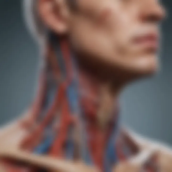
Understanding the vascular structures in the head and neck is crucial for grasping the interconnectivity and functionality of this complex region. These structures are not just conduits for blood but also play vital roles in maintaining homeostasis and providing essential nutrients to the tissues. The arteries, veins, and lymphatics create a network that supports not only the physiological but also the pathological aspects of human health. It’s these very intricacies that highlight the importance of studying vascular anatomy for anyone involved in healthcare and related fields.
Major Arteries
The head and neck are supplied with blood primarily by the carotid and vertebral arteries. The common carotid artery bifurcates into the internal and external carotid arteries, establishing a vital supply line to the brain and face.
- Internal Carotid Artery: This artery ascends the neck to provide blood to the brain. It branches into several key arteries, including the ophthalmic artery which supplies the eyes, and the middle cerebral artery, crucial for many brain functions.
- External Carotid Artery: This artery’s branches serve the face and neck. Key branches include the facial artery, which supplies the skin and muscles of the face, and the maxillary artery, essential for deep facial structures, including parts of the jaw and teeth.
The significance of understanding these arteries cannot be overstated. Any occlusion or injury can lead to serious complications, including strokes or facial ischemia.
Veins and Venous Drainage
Veins in the head and neck carry deoxygenated blood back to the heart. Major veins include the jugular veins. The arrangement and function of these veins highlight the unique considerations of blood drainage in this area.
- Internal Jugular Vein: This vein drains blood from significant regions of the brain, the face, and most areas of the neck. It forms part of a complex drainage system that also works in tandem with arterial pathways to ensure efficient circulation.
- External Jugular Vein: More superficial, this vein drains the areas served by the external carotid artery, such as the scalp and outer face. Understanding the drainage patterns of these veins is essential, especially for procedures that may risk compromising these structures.
- Cavernous Sinus: An important clinical consideration, this sinus collects blood from the ocular and cerebral regions. It communicates with the internal jugular vein, establishing a crucial route for venous drainage but also a potential pathway for infectious spread, which can have dire consequences.
Lymphatic System
The lymphatic system in the head and neck plays a pivotal role in immune response, tissue fluid balance, and absorption of fats. It's a network that can seem quite overwhelming but is essential for good health.
- Lymph Nodes: Numerous lymph nodes situated throughout the neck help filter lymph fluid, providing a first line of defense against pathogens. The cervical lymph nodes are particularly significant as they respond to infections or malignancies, where they may become enlarged or tender.
- Lymphatic Vessels: These vessels transport the lymph fluid away, connecting to the larger lymph ducts. They are integral in the absorption of extracellular fluid and its return to the bloodstream, which is crucial in maintaining fluid balance in the body.
- Thoracic Duct: This is the largest lymphatic vessel, draining lymph from the majority of the body, including the left side of the head and neck. It plays a vital role in the overall lymphatic system by returning lymph to the circulation, ensuring that nutrients and waste products are efficiently processed.
The lymphatic system is not only vital for immune functions but also plays significant roles in fluid regulation and fat absorption.
In summary, the vascular structures of the head and neck are foundational to health and disease management. Their comprehension is essential for clinicians, educators, and students alike, providing critical insights into both physiological processes and potential pathologies.
Organs in the Head and Neck
Understanding the organs in the head and neck is crucial for several reasons. This region is not only the control center for many vital functions but also houses structures essential for communication, metabolism, and overall homeostasis. The interplay of these organs affects daily life in profound ways, from the way we speak and eat to how we express emotions and respond to our environment.
Salivary Glands
Salivary glands are significant players in both digestion and oral health. They produce saliva, which moistens food, making it easier to swallow and start the digestive process. Saliva contains enzymes like amylase that begin breaking down carbohydrates even before food reaches the stomach. Additionally, saliva helps wash away food particles, minimizing the risk of tooth decay and gum disease.
There are three major pairs of salivary glands: the parotid glands, submandibular glands, and sublingual glands. Each varies in size and function:
- Parotid glands: The largest, located near the cheek, they produce a serous, watery secretion that contains significant amounts of amylase.
- Submandibular glands: These are found under the jaw and produce both serous and mucous secretions.
- Sublingual glands: The smallest, situated beneath the tongue, they primarily secrete mucous.
Understanding how these glands function aids in recognizing conditions like xerostomia, commonly known as dry mouth, which can lead to eating difficulties and dental problems.
Thyroid and Parathyroid Glands
The thyroid and parathyroid glands play pivotal roles in regulating metabolism and calcium levels, respectively. The thyroid gland, shaped like a butterfly, is situated at the base of the neck and produces hormones including thyroxine (T4) and triiodothyronine (T3). These hormones govern metabolism, influencing how the body converts food into energy. Insufficient hormone production can lead to conditions such as hypothyroidism, characterized by fatigue and weight gain, while excessive hormone production can result in hyperthyroidism, leading to symptoms like anxiety and weight loss.
The parathyroid glands, typically four in number, are small and located adjacent to the thyroid. They secrete parathyroid hormone (PTH), crucial for maintaining calcium homeostasis. PTH regulates calcium levels in the blood, stimulating the release of calcium from bones and increasing absorption from the intestine. Disturbances in this balance can lead to disorders like hyperparathyroidism or hypoparathyroidism, with direct implications for bone health.
Larynx and Its Functions
The larynx, commonly called the voice box, sits at the junction of the pharynx and trachea. Its primary functions are vital and multifaceted. During breathing, the larynx allows air to pass into the trachea, and in the act of swallowing, it prevents food from entering the respiratory tract, protecting the airway.
Its most notable feature is its role in phonation. The laryngeal muscles adjust the tension of the vocal cords, modulating pitch and volume when we speak or sing. The anatomy of the larynx includes several important structures:
- Epiglottis: A flap that closes over the larynx during swallowing.
- Vocal cords: These are folds of tissue that vibrate to produce sound.
- Laryngeal cartilages: These provide structure and support, crucial for maintaining an open airway.
Issues with the larynx can lead to voice disorders, which can significantly affect communication. Understanding its anatomy aids in diagnosing conditions such as laryngitis or vocal cord nodules.
The organs in the head and neck not only serve individual functions but are interconnected, influencing both health and quality of life in myriad ways.
The Respiratory System
The respiratory system is more than just a series of tubes and sacs; it's the lifeline that fuels our bodies with oxygen and expels carbon dioxide. Within the context of head and neck anatomy, understanding this system is crucial, as it interlinks with several anatomical structures and functions. The respiratory system not only facilitates breathing but also plays a vital role in phonation, olfaction, and protecting the respiratory tract from pathogens. A well-functioning respiratory system is essential for overall health, while disorders can impact the quality of life significantly.
Anatomy of the Nasal Cavity
The nasal cavity is the gateway to the respiratory system. It lies just behind the nose and serves several key functions. Primarily, it filters, warms, and humidifies the air inhaled before it travels to the lungs. The anatomy of the nasal cavity includes the nasal septum, which divides it into two halves, and the intricate network of conchae (turbinate bones) that increases the surface area. These structures are lined with a mucous membrane that helps trap dust and microbes, reducing the chance of respiratory infections.
- Functions of the Nasal Cavity:
- Filtration: Prevents particulate matter from entering the lungs.
- Conditioning: Warms and moistens the air, making it less harsh on the bronchial tree.
- Olfaction: Houses millions of olfactory receptors for the sense of smell.
In addition, the nasal cavity connects to the sinuses, which also play a role in humidifying the air and lightening the skull. Sinus infections can lead to pressure and discomfort, showcasing just how interconnected these structures are.
Pharynx and Its Role
The pharynx is a muscular tube that acts as a passageway for both air and food. It connects the nasal cavity and mouth to the larynx and esophagus, respectively. The pharynx is divided into three sections: nasopharynx, oropharynx, and laryngopharynx. Each section has its specific roles in respiratory and digestive functions.
- Sections of the Pharynx:
- Nasopharynx: Located behind the nasal cavity, it serves as an air passage.
- Oropharynx: The part behind the mouth that serves as a common pathway for food and air.
- Laryngopharynx: The lower part that directs air into the larynx and food into the esophagus.
The pharynx is equipped with muscles that facilitate swallowing and prevent food from entering the airways. A reflex action prompts the epiglottis to close off the larynx while swallowing, demonstrating how these anatomical features work in harmony.


“The pharynx symbolizes the convergence of the respiratory and digestive pathways, playing a critical role in human survival.”
Trachea and Bronchial Tree
The trachea, commonly known as the windpipe, extends from the larynx and branches into the right and left bronchi which lead to each lung. The trachea is reinforced with C-shaped rings of cartilage that keep it open during breathing, preventing collapse. It is lined with ciliated epithelium that traps and propels mucus and dust away from the lungs.
The bronchial tree represents the branching structure of airways within the lungs, further dividing into smaller bronchi and bronchioles. This extensive network facilitates efficient gas exchange at the alveolar level.
- Key Points about the Trachea and Bronchial Tree:
- C-Shape Cartilage: Ensures air passage remains open.
- Mucociliary Escalator: A defense mechanism that clears mucus and debris.
- Alveoli: Tiny air sacs at the end of the bronchioles where oxygen and carbon dioxide exchange occurs.
Understanding the intricacies of the respiratory system's layout helps illustrate its vital role in both everyday life and in medical pathology.
By comprehending these anatomical landmarks and functions, medical professionals and students can better appreciate how disruptions in this system can significantly affect health and well-being, emphasizing its importance in clinical practice.
Dental Anatomy
Understanding dental anatomy is crucial in the broader landscape of head and neck anatomy. This field focuses primarily on the structure and function of teeth and their supporting tissues. Within this article, dental anatomy is presented as an integral component that not only affects oral health but also has implications for overall systemic health. The mouth is often viewed as the gateway to the body, and the state of one’s dental structures can significantly inform medical professionals about a patient’s general condition.
Several specific elements of dental anatomy deserve attention. First and foremost, the structure of the tooth itself allows us to appreciate how each component plays a role in the processes of chewing and, by extension, digestion. Additionally, different types of teeth are tailored for distinct functions, contributing to the intricate ballet of oral mechanics that we often take for granted. The relationship between teeth and the gingiva, or gums, along with other periodontal structures, cannot be overlooked; these elements work together to maintain both function and aesthetic appeal—a vital consideration in dental practice.
In summary, dental anatomy is not merely an isolated topic but interacts deeply with various anatomical and physiological processes. Leveraging knowledge within this area can help in various professional arenas, including dentistry, surgery, and holistic medicine.
Structure of a Tooth
The structure of a tooth can be divided into several specific layers, each with defined roles. At the very core lies the pulp, a soft tissue housing nerves and blood vessels, essential for the vitality of the tooth.
- Dentin – This layer surrounds the pulp and provides support. Comprised of mineralized tissue, dentin is crucial for the strength of the tooth.
- Enamel – The hardest substance in the human body, enamel covers the outer surface of the tooth and protects the underlying dentin and pulp from damage and decay.
- Cementum – This layer helps anchor the tooth in its socket and assists in periodontal health.
Together, these components form a robust structure designed to withstand the daily rigors of biting and chewing. Each component possesses unique properties that contribute to the health and longevity of teeth.
Types of Teeth and Their Functions
Teeth are categorized into different types, each serving specific functions within the oral cavity. Understanding these types helps clarify their roles in eating and overall oral function.
- Incisors: These are the front teeth, sharp and chisel-shaped, designed for slicing food. There are four incisors in each quadrant of the mouth.
- Canines: Pointed and prominent, canines are adept at tearing food. Their position in the mouth lends them strength.
- Premolars: These teeth, located behind the canines, have a flat surface for crushing and grinding food, making them indispensable for proper mastication.
- Molars: The larger teeth at the back of the mouth, designed for grinding and chewing. Their broad, flat surfaces help break down food into smaller particles.
In summary, each type has developed evolutionary traits aimed at optimizing food intake, contributing to the overall efficiency of the digestive process.
Gingiva and Periodontal Structures
The gingiva, the part of the gum around the base of each tooth, plays a significant role in oral health. Healthy gums form a tight seal around the teeth, protecting the underlying bone and supporting structures from infection.
Periodontal structures refer to the supporting tissues that include:
- Periodontal Ligament: This connective tissue anchors the tooth to the bone, allowing for slight movement which is critical for absorbing biting forces.
- Alveolar Bone: The bone that houses the tooth roots provides support and stability. It's essential that this bone remains healthy to prevent tooth loss.
A consideration of these structures reveals their importance in diagnosing and treating various dental diseases. Periodontal disease, for example, can lead to systemic complications, making an understanding of these structures essential for healthcare professionals focusing on the head and neck anatomy.
"Tooth health reflects overall health, intertwining systemic well-being with oral hygiene practices."
Integumentary System
The integumentary system serves as the body’s primary interface with its environment, encompassing not just the skin but also hair follicles, glands, and connective tissues that contribute to overall health and functionality. Within the context of head and neck anatomy, understanding this system is crucial for both medical professionals and students. It provides insights into conditions that may arise in this region and informs various clinical practices. The skin acts as a barrier, while hair and glands play essential roles in thermoregulation and sensation. This section will dive deep into these components, demonstrating their importance in maintaining homeostasis and protecting underlying structures.
Skin Anatomy of the Face and Neck
The skin covering the face and neck has unique characteristics that distinguish it from the skin of other body parts. For one, it is generally thinner and more sensitive, which can make it susceptible to injury and various dermatological conditions.
- Epidermis: The outermost layer, responsible for protection and the first line of defense against environmental hazards. The epidermis here is rich in melanocytes, which give skin color and provide some shield against UV radiation.
- Dermis: This thicker layer contains collagen and elastic fibers, providing flexibility and strength. The dermis also houses most of the skin's appendages, including hair follicles and glands.
- Subcutaneous Tissue: This layer connects the skin to underlying structures like muscles and bones. It contains fat, which not only acts as an insulator and shock absorber but also as an energy reservoir.
Understanding these layers contributes significantly to both aesthetic and medical applications. For example, during surgical procedures like facelifts or skin grafts, knowledge of the skin’s anatomy is paramount to minimize complications and promote healing.
Hair Follicles and Glands
Hair follicles and glands intertwined with the skin play vital roles in maintaining homeostasis. The follicles are responsible for producing hair, which serves not just a sensory purpose but also helps with thermoregulation. Each follicle is associated with sebaceous glands that secrete sebum, an oily substance that helps to keep skin moisturized and acts as a barrier against microbes.
- Types of Hair: On the face and neck, you'll find different types, from fine, vellus hair to thick terminal hair, such as beard hair. This distinction is not merely cosmetic; it has implications in endocrinology, as hair type can be influenced by hormones.
- Gland Functions: Apart from sebaceous glands, you also have sweat glands, which are essential for thermoregulation by allowing the body to cool through perspiration.
The dysfunction of these glands can lead to conditions such as acne or eczema, showcasing the need to understand their anatomy to develop effective treatments.
Connective Tissue Layers
Beneath the skin, the connective tissue layers serve critical functions, ensuring structural integrity and supporting the skin and underlying organs. These layers are not uniform and vary across different areas of the face and neck, impacting how injuries heal and how surgical interventions should be approached.
- Composition: The connective tissue is comprised of various cell types, fibers, and ground substance. Collagen provides tensile strength, while elastin contributes elasticity, allowing the skin to stretch and retract.
- Fascial Layers: The fascia in the head and neck can be divided into several layers, playing crucial roles in compartmentalization and in aiding the movement of muscles and nerves. Understanding this anatomy is essential for approaches in procedures like facelifts or reconstructive surgeries.
Functional Considerations
When discussing the anatomy of the head and neck, it's crucial to highlight the functional considerations that underpin various activities we often take for granted. This section dives deep into how the structures within this region work synergistically to facilitate critical functions such as facial expression, speech, and swallowing. Understanding these aspects not only bridges theoretical knowledge with practical implications but also emphasizes the relevance of anatomical studies in addressing real-world clinical issues.
Facial Expression Mechanisms
Facial expressions are not merely aesthetic; they play a vital role in non-verbal communication, emotional expression, and even social interaction. At the heart of this phenomenon are the muscles of facial expression, primarily innervated by the facial nerve, also known as cranial nerve VII. These muscles coordinate to produce a vast range of expressions, from joy to sorrow, facilitating a rich tapestry of human interaction.
For instance, consider the use of the orbicularis oculi – the muscle encircling the eye. This muscle allows one to tightly close the eyelids, essential for protecting the eyes and conveying emotions like surprise or happiness. The zygomaticus major, another key player, helps lift the corners of the mouth, essential for smiling.
The ability to convey emotions through facial mechanics is not just an anatomical curiosity; it holds substantial implications in fields like psychology, medicine, and art therapy. For those studying conditions that affect facial muscles, like Bell's palsy, understanding these mechanisms aids in developing effective rehabilitation strategies.
Speech and Phonation
The structures of the head and neck are crucial in the production of speech and phonation. The larynx, or voice box, serves as the primary organ responsible for sound production, housing the vocal cords. When air is pushed from the lungs through the trachea, it passes through the larynx; the vibrations of the vocal cords create sound waves, which are then modified by the resonating chambers of the throat, mouth, and nasal passages.
Articulation involves the precise movements of the tongue, lips, and palate. For example, the tensor veli palatini and levator veli palatini muscles adjust the soft palate's position, enabling us to switch between nasal and oral sounds. This intricate coordination is essential for producing intelligible speech, and any disruption—due to disease or injury—can lead to significant communication challenges.
Moreover, the study of speech mechanics intersects with various disciplines, from linguistics to speech pathology, demonstrating the multifaceted applications of functional anatomy in addressing disorders like stuttering or dysarthria.
Swallowing Mechanisms
Swallowing, a seemingly simple act, involves a complex series of coordinated movements that engage various anatomical structures. This process can be categorized into three phases: oral, pharyngeal, and esophageal. At the heart of swallowing is the synergy between the muscles of the floor of the mouth, the pharynx, and the esophagus.
During the oral phase, food is manipulated and formed into a bolus by the tongue. The muscles involved include the genioglossus and hyoglossus, which help shape the food for easier swallowing. Once the bolus is ready, it moves into the pharyngeal phase, where the soft palate elevates, closing off the nasal passage to prevent aspiration. Simultaneously, the larynx moves upward, and the epiglottis folds down to protect the airway.
The final esophageal phase involves a series of peristaltic movements that push the bolus into the stomach. Any disruption in this finely tuned process can lead to issues ranging from choking to aspiration pneumonia. Hence, understanding the anatomy involved in swallowing not only enriches our knowledge but also informs clinical practices concerning dysphagia management.
"The intricate relationship of anatomical structures in the head and neck illustrates how essential they are in our daily functions, impacting both health and quality of life."
Clinical Relevance
The study of head and neck anatomy holds paramount significance in various fields such as medicine, dentistry, and allied health. This intricate area of the human body is not only rich in complex structures but also plays pivotal roles in vital functions, including respiration, digestion, communication, and sensory perception. Understanding the clinical relevance of these anatomical features enhances the diagnosis and treatment of various conditions.
For practitioners, knowledge of head and neck anatomy is essential for assessing injuries, detecting diseases, and developing treatment plans. Mastery of the anatomical landscape allows healthcare professionals to accurately interpret symptoms and identify underlying health issues, potentially improving patient outcomes. Key benefits of this knowledge include:
- Enhanced diagnostic capabilities
- Informed treatment choices
- Optimal surgical planning and execution
Moreover, the interconnections between various structures in this region underline the importance of multimodal learning, incorporating clinical correlations to foster a deeper understanding. For instance, conditions like TMJ dysfunction and sleep apnea directly link to anatomical variations in the jaw and airway structures. Recognizing these relationships aids in tailoring individual management strategies.
Understanding the anatomical intricacies is key for effective clinical practice, ensuring healthcare providers can address both common and complex issues with confidence.
Common Disorders and Conditions
Various disorders and conditions can affect the structures within the head and neck region. A deep understanding of the anatomy assists in the identification and management of these ailments. Some prevalent disorders include:
- Sinusitis: Inflammation of the sinuses often caused by infection, leading to facial pain and nasal congestion.
- Temporomandibular Joint Disorders (TMJ): Conditions affecting the jaw joint, resulting in pain, clicking, and difficulty in jaw movements.
- Thyroid Disorders: Including hypothyroidism and hyperthyroidism, these conditions impact metabolic functions and may lead to significant systemic effects.
- Oral Diseases: Such as periodontal disease, which affects gums and teeth, impacting overall health and necessitating proper dental care.
Recognizing these disorders hinges on a comprehensive grasp of the underlying anatomical structures, allowing for targeted interventions and management.
Diagnostic Techniques
Diagnosing conditions pertaining to the head and neck often requires a blend of clinical examination and advanced imaging techniques. These diagnostic tools are essential for visualizing structures and identifying abnormalities. Common techniques include:
- Radiography: X-rays highlight bony structures and can be pivotal in detecting fractures or dental issues.
- CT Scans: Computed Tomography provides detailed cross-sectional images and is invaluable for assessing complex craniofacial skeletons, often utilized in trauma cases.
- MRI: Magnetic Resonance Imaging is excellent for visualizing soft tissues, making it useful for evaluating neurological, vascular, and muscular conditions.
- Ultrasound: Sometimes used in assessing thyroid nodules or lymph nodes, ultrasound can guide biopsies and other procedures.
Each diagnostic method possesses unique strengths, guiding healthcare providers toward appropriate treatment decisions.
Surgical Interventions
Surgical approaches in the head and neck region are diverse, reflecting the complexity of the anatomy and common pathologies encountered. Procedures may range from minor to extensive, emphasizing the need for a detailed understanding of the anatomy to minimize complications. Notable surgical interventions include:
- Thyroidectomy: The removal of thyroid tissue, often necessitated by the presence of tumors or hyperthyroidism.
- Laryngectomy: Involves partial or total removal of the larynx, commonly due to malignancies, impacting speech and airway.
- Jaw surgery: Such as orthognathic surgery, to correct jaw alignment and improve bite function.
- Facial reconstructive surgery: Aimed at restoring appearance and function following trauma or congenital defects.
Each surgical intervention underscores the intricate relationship between anatomy and patient care, where knowledge translates into improved surgical outcomes and patient satisfaction.
Imaging Techniques in Anatomy
Imaging techniques play a pivotal role in understanding the anatomy of the head and neck. Given the complexity and nuances involved in this region, such techniques provide invaluable insights that can bridge theoretical knowledge with practical application. With the rapid advancement in technology, imaging not only serves as a diagnostic tool but also aids in research, education, and surgical planning.
Importance of Imaging Techniques
- Enhanced Visualization: Imaging provides clarity that is often unachievable through traditional methods. It reveals the layers and structures within the head and neck, helping healthcare professionals visualize intricate details that may affect diagnosis and treatment.
- Non-Invasive Methods: Techniques like MRI or CT are non-invasive and allow for treatment planning without the need for surgical intervention. This significantly reduces patient risk and enhances overall treatment protocols.
- Guiding Surgical Interventions: Surgeons rely on imaging to map out the anatomy before performing complex procedures. Having a visual guide can delineate critical structures, ensuring safety and efficacy.
- Assessment of Conditions: Imaging techniques are essential in identifying anomalies, tumors, or injuries within the head and neck region. They enable practitioners to make accurate assessments that can lead to timely and appropriate interventions.
Overall, imaging techniques are not just tools; they are essential components of modern medical practice, refining our understanding and management of anatomical structures.
Radiography
Radiography is one of the oldest imaging techniques and continues to be a first-line tool in examining the head and neck. X-ray technology utilizes ionizing radiation to create images of structures. In the context of the head and neck, it can be particularly useful for evaluating bony structures and detecting fractures.
- Benefits:
- Considerations:
- Quick acquisition of images enables immediate decisions in emergency settings.
- Provides a broad overview of bone integrity and alignment.
- Limits include less resolution for soft tissue assessment; thus, it cannot replace more advanced modalities when soft tissue detail is necessary.
Generally, while radiography excels in assessing skeletal anatomy, it is vital to consider its limitations relative to other imaging techniques.
Radiographs can also assist in monitoring dental health and conditions affecting the salivary glands, crucial for a comprehensive understanding of head and neck anatomy.
MRI and CT Imaging
MRI (Magnetic Resonance Imaging) and CT (Computed Tomography) imaging have emerged as powerful tools for detailed anatomical visualization.
- MRI: This non-invasive method utilizes magnetic fields and radio waves to produce highly detailed images of soft tissues. With exceptional contrast resolution, MRI is outstanding for studying complex tissues, such as
- CT Scanning: CT imaging combines X-ray technology with computer processing to create cross-sectional images of the body. It is particularly beneficial for identifying
- The brain
- Muscles
- Nerves and Blood vessels
- Bone fractures
- Tumor locations and sizes
Both MRI and CT imaging have their unique relevance depending on the clinical scenario. The former excels in soft tissue differentiation, while the latter offers faster imaging for acute conditions. In many cases, they can be complementary, enhancing the overall diagnostic process.
To conclude, mastering imaging techniques is not merely a technical skill but a fundamental aspect of effective anatomical understanding. Within this intricate landscape of the head and neck, these imaging modalities illuminate the shadows, revealing both structure and function.
Future Trends in Anatomical Research
The exploration of anatomical research is continuously evolving, shedding light on areas that enhance both education and practical medical applications. In this section, we will explore key advancements that represent the cutting edge of our understanding of head and neck anatomy as well as discuss the broader implications for clinical practice. Programs utilizing innovative techniques can provide clearer insights into the complexities of anatomy, offering paths to improve patient care and outcomes.
Advancements in Technology
Modern anatomical research is experiencing a technological renaissance. The marriage between technology and anatomy opens doors that were previously too intricate or fraught with challenges to navigate. Here are some of the notable advancements:
- 3D Printing: The ability to create detailed, life-sized models of anatomical structures allows for unprecedented study and interaction. Surgeons can rehearse operations on these printed models, leading to more precise and thoughtful interventions.
- Virtual Reality: VR technology is redefining the learning and teaching of anatomy. With immersive simulations, students and professionals alike can visualize and manipulate anatomical structures in three-dimensional space, significantly enhancing their understanding.
- Augmented Reality: This technology overlays digital information on physical structures, providing a richer context for learning. Imagine being able to see the blood flow in real-time as you examine the cardiovascular aspects of the anatomy.
- Artificial Intelligence: AI is aiding researchers in recognizing patterns and anomalies in anatomical scans, delivering precise interpretations, and even predicting surgical outcomes.
These advancements not only facilitate enhanced learning but also promote deeper investigations into complex anatomical relationships.
Implications for Medical Practice
As with any advancement, new technologies in anatomical research carry significant implications for medical practice. The integration of these innovations has the potential to improve clinical outcomes in several nuanced ways:
- Enhanced Surgical Precision: By utilizing models from 3D prints or VR simulations, surgeons can approach procedures with greater confidence, as they can visualize the anatomy they are interacting with before making any incision.
- Personalized Medicine: With technology enabling more intricate understanding of individual anatomical variations, treatments can be tailored more closely to the patient's specific needs, driving down instances of surgical failure and complications.
- Improved Training for Medical Professionals: The aforementioned technologies serve not only as tools for surgeons but also for education, helping to cultivate more competent and confident healthcare providers.
- Interdisciplinary Collaboration: These advancements promote interactions between surgeons, radiologists, and anatomical researchers, fostering a multidisciplinary approach that enhances patient care through combined expertise.
"In the rapidly evolving landscape of medical science, those who adapt will thrive. The integration of anatomy into technological advancements is no longer future talk; it’s our reality."
The End
In the intricate tapestry of head and neck anatomy, understanding the core elements is paramount not only for medical professionals but also for anyone with a keen interest in human biology. The conclusion serves as a pivotal point, weaving the detailed threads of knowledge explored throughout this article into a cohesive understanding. This section emphasizes the importance of synthesizing the complex information presented, allowing both students and researchers to grasp the relationships and functions within this fascinating region of the body.
Through our exploration, key elements such as the relationship between the cranial and facial bones, the multifaceted roles that muscles play in daily activities, and the various systems at work—like vascular and nervous—are highlighted. These insights not only reinforce the critical nature of anatomical knowledge but also bridge the gap between theory and practical application, which is often crucial in medical contexts.
The benefits of comprehending head and neck anatomy extend well beyond academia. For clinicians, it provides a foundation for diagnosing and treating conditions effectively. Even for those in related fields, the knowledge fosters a greater appreciation for the intricate design of the human body and its capabilities.
Summary of Key Points
- Cranial and Facial Bones: Their structure and function lay the groundwork for understanding facial features and protective roles.
- Muscular Interactions: The muscles of the face and neck facilitate not just movement but also vital functions such as swallowing and articulation.
- Vascular and Nervous Systems: These systems play crucial roles in supplying oxygen and nutrients as well as in controlling involuntary and voluntary movements.
By revisiting these points, we ensure clarity in the comprehension of how these various aspects interlink, contributing to the overall functionality of the head and neck.
Final Thoughts
In closing, this article serves as a crucial resource for anyone delving into the realm of head and neck anatomy. The depth of information provided aids significantly in advancing knowledge and expertise. As we move forward in medical research and practice, the exploration of this complex anatomy will no doubt continue to evolve, reminding us of the continuous journey of learning and discovery. Whether you're a seasoned professional or a student just beginning your studies, the content within this article offers invaluable insight that will serve you as a solid foundation in understanding one of the most intricate areas of human anatomy.


